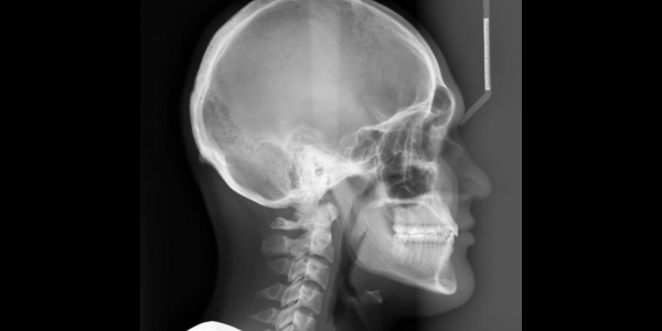
Lateral Cephalogram

A Lateral Cephalogram (or Lat Ceph) is an x-ray taken of the side of the face with very precise positioning so that various measurements can be made to determine the current and future relationship of the top and bottom jaw (maxilla and mandible) and therefore assess the nature of a patient’s bite. This is particularly useful to plan any orthodontic treatment that may be necessary.

As treatment progresses, it is helpful to have any future Lateral Cephalograms taken at the same practice so that the x-rays can be easily compared.

Frontal Cephalogram (P-A Cephalogram)
A frontal cephalogram is a radiograph of the head taken with the x-ray beam perpendicular to the patient’s coronal plane with the x-ray source behind the head and the film cassette in front of the patient’s face. P-A cephalograms are usually taken for evaluation and treatment planning of patients with facial asymmetry.
Both Temporomandibular Joint (Both TMJ)

Temporo-mandibular joint (TMJ) disorders are medical problems related to the jaw joint, which connects the jaw or mandible to the temporal bone at each side of the head. As you open and close your mouth each time you talk, chew, sing or yawn, your jaw condyles glide along the joint socket of the temporal bone.

Standard x-rays of the TMJs are taken in the open and closed position for each side. Additional, advanced medical imaging, such as Cone Beam CT (CBCT) might also be needed to fully assess the TMJs.

Frequent QnA

During the OPG the arm of the machine will rotate slowly around the head but will not come into contact with the patient. The Radiographer (a technologist trained in medical and dental imaging) will instruct the patient to bite on a small plastic mouthpiece attached to the machine, which keeps the top and bottom teeth separated and helps position the mouth properly in the machine.

For a Lateral Cephalogram you are required to hold still and bite together on the back teeth. Lips should be relaxed. The Radiographer will help ensure that patients are in the correct position before taking the x-ray.

The procedure is very brief (about 5 minutes or less) and is painless and simple. It is very important that you follow all instructions and hold absolutely still once positioned.
There is no special preparation required and there are no after effects following the scan. Once at the practice, patients will be asked to remove any jewellery or metallic items from the head and neck region, including any piercings, hair accessories, dentures and plates, as these often interfere with x-ray image formation and produce unwanted artefacts.